Andor Zyla sCMOS enables breakthrough in neuronal activity research
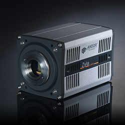
Photo by Andor Technology plc.
The speed, resolution and sensitivity of the Andor Zyla sCMOS camera has allowed the Vaziri research group in Vienna, Austria, to simultaneously image neuronal activity across an entire organism for the first time. This profound advance follows closely on their innovative wide-field temporal focusing (WF-TeFo) two-photon approach to high-speed imaging of whole brains, which was also enabled by an Andor camera – the Neo sCMOS.
Publishing in Nature Methods, the team describe using their new technique of light-field deconvolution microscopy (LFDM) to map the simultaneous neuronal population activity of a whole nematode worm, Caenorhabditis elegans, as well as the whole-brain of zebrafish larvae in combination with sensory stimulation 20x faster than previously achieved using other techniques.
According to lead author, Dr Robert Prevedel, “LFDM elegantly combines light-field microscopy with 3D deconvolution and enables us to image a whole 3D-volume with a single exposure to a 2D sensor. This simultaneous 3D imaging is scan-less and involves no moving parts during acquisition, can be easily added to any commercial microscope, and is very cost-effective.
“Our experiments on whole organism imaging demonstrate the sheer power of LFDM for high-speed imaging, which is limited only by the camera frame-rate and the signal strength of the Ca-reporter. Our nuclear-confined Calcium-indicator, which visualises the activity of a neuron by increasing its fluorescence, was combined with the ultra-fast frame rate of the Andor Zyla 5.5 megapixel sCMOS camera for unambiguous discrimination of individual neurons across C. elegans. The 100 Hz full-frame Zyla camera was one of the two fastest sCMOS cameras available and in the development of our WF-TeFo microscopy technique we had been very satisfied with the performance of its predecessor, the 30 Hz Andor Neo.”
Dr Prevedel describes the performance of the Zyla sCMOS camera as “flawless” and continues: “We especially liked the capability of the control software, SOLIS, to capture, stream and display the huge image data in real time and with minimal efforts. This way we could watch the data acquisition in real time and effortlessly browse through many GBs of recordings. In the end, we only needed to deploy the Zyla at 50 Hz but the extra speed capability may be very useful in future experiments.”
Although light-field microscopy (LFM) has been successfully applied to imaging still and in-vitro biological samples, it has not been used for any functional biological imaging. This is due largely to its reduced spatial resolution compared to standard microscopy methods and the inherent trade-off between the spatial and axial resolution as well as axial imaging range. LFDM combines LFM with a 3D deconvolution algorithm whereby to achieve an effective resolution of approx. 1.4 μm and 2.6 μm in the lateral and axial dimensions respectively inside biological samples using a 40x objective while simultaneously recording neuronal population activity over a field of view of approx. 350 μm x 350 μm x 30 μm. This is sufficient to capture the dynamics of neurons distributed across the entire nervous system of C. elegans.
Using a 20x 0.5NA objective, the group also applied their imaging technique to capture the neuronal activity of zebrafish whole brains, were they achieved instantaneous imaging of massive field-of-views of 750 μm x 750 μm x 200 μm with spatial resolution of approx. 3.5 μm laterally and 11 μm axially.
“The value of this work to the neuroscience community is evident in the immediate attention it attracted,” says Orla Hanrahan of Andor. “And, because their imaging methodology can be applied quite simply and straightforwardly to existing microscopes, it should find immediate and widespread use.”
Unlike previous CMOS or CCD technologies, the Andor Zyla 5.5 sets radical new benchmarks in its unique ability to simultaneously deliver highest specifications in sensitivity, resolution, speed, dynamic range and field-of-view. The camera is based around a large 5.5 megapixel sensor with 6.5 µm pixels and a 22 mm diameter and is capable of 100 fps sustained (Camera Link; 40 fps, USB3), making it ideal for applications in cell microscopy, astronomy, digital pathology, and high content screening. The Rolling and Global shutter exposure modes further enhances application flexibility. Global shutter in particular offers an ideal means to simply and efficiently synchronize the Zyla with other ‘moving’ devices, such as stages or light switching sources, and eliminating the possibility of spatial distortion when imaging fast moving objects.
For more information, please visit http://www.andor.com.
News Categories
- » NEWS HOME
- » Automation & Robotics
- » Industry 4.0
- » Material Handling
- » Sensors
- » Quality & Testing
- » Machine Vision
- » Laser & Optics
- » Metalworking
- » Motion Control & Drives
- » Hydraulics & Pneumatics
- » Process Industry
- » Renewable Energy
- » Agriculture
- » Home & Office Furniture
- » Environmental Tech

Tags
|
|
|
|
|
|
|
|
|
|
|
|
|
|
|
|
|
|