Andor Neo camera contributes to breakthrough in brain-wide imaging
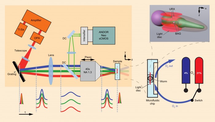
Photo by Andor Technology plc.
Although the entire nervous system connectivity map of the nematode model organism, Caenorhabditis elegans, has been known for more than 25 years, it has not been possible to predict all functional connections underlying behaviour. Now, the Vaziri and Zimmer research groups in Vienna, Austria, have demonstrated a high-speed imaging technique that allows neuronal activity across the entire brain to be recorded simultaneously.
Publishing in Nature Methods, Professor Alipasha Vaziri ascribes two key breakthroughs for their success in being the first to implement brain-wide imaging. “By choosing the Andor Neo 5.5 megapixel sCMOS camera, we were able to image the entire volume associated with the worm’s brain (75 x 75 x 30 μm) four to six times per second using wide-field temporal focusing (WF-TeFo) two-photon microscopy. Plus, we used a novel Calcium-indicator that visualises the activity of a neuron by increasing its fluorescence and permits unambiguous discrimination of individual neurons within the densely packed head ganglia.”
Dr Manuel Zimmer backs this up, saying “The Andor Neo camera was one of the only two sCMOS cameras then available and our demo-trial convinced us that it provided the fastest speed while retaining high sensitivity and a wide dynamic range. We also chose Andor over its competitors because of its capability to integrate easily into our home-made hardware control interface, as well as their good customer and technical support service.”
WF-TeFo microscopy allows users to independently control the axial and lateral confinement of the excitation area while taking advantage of the high depth penetration and low scattering properties of two-photon excitation. For their volumetric imaging, the group scanned an excitation area of approx. 60 μm in diameter with an axial confinement of approx. 1.9 μm in the axial direction only to speed up the volume acquisition time. A volume acquisition rate approaching 13 Hz was achievable but imaging was performed at speeds of 4–6 Hz, corresponding to approx. 5.6 megavoxels per second, achieving a lateral spatial resolution of 0.285 μm.
“The value of this work to the neuroscience community is evident in the attention it attracted online, ranking second overall for Nature Methods,” says Orla Hanrahan of Andor. “Because their imaging methodology can also be applied straightforwardly to other organisms, or more general imaging tasks, and since their novel calcium reporter is a very powerful tool that can be used by all neuroscientists working on C. elegans, their technique has already had a large impact. If a lab-on-a-chip device is deployed for stimulus delivery in the future, this method provides an enabling platform for establishing functional maps of neuronal networks.”
Unlike previous CMOS or CCD technologies, the Andor Neo 5.5 sets radical new benchmarks in its unique ability to simultaneously deliver highest specifications in sensitivity, resolution, speed, dynamic range and field-of-view. The camera is based around a large 5.5 megapixel sensor with 6.5 µm pixels and a 22 mm diameter and is capable of 30 fps sustained or up to 100 fps burst to the 4 GB internal memory, making it ideal for applications in cell microscopy, astronomy, digital pathology, and high content screening. Built on a -40°C vacuum-cooled platform, the extremely low darkcurrent and 1 e- read noise means Neo 5.5 is suited to a range of exposure conditions. The Rolling and Global shutter flexibility further enhances application flexibility, Global shutter in particular offering an ideal means to simply and efficiently synchronize the Neo with other ‘moving’ devices, such as stages or light switching sources, and eliminating the possibility of spatial distortion when imaging fast moving objects.
FOr more information, please visit http://www.andor.com.
News Categories
- » NEWS HOME
- » Automation & Robotics
- » Industry 4.0
- » Material Handling
- » Sensors
- » Quality & Testing
- » Machine Vision
- » Laser & Optics
- » Metalworking
- » Motion Control & Drives
- » Hydraulics & Pneumatics
- » Process Industry
- » Renewable Energy
- » Agriculture
- » Home & Office Furniture
- » Environmental Tech

Tags
|
|
|
|
|
|
|
|
|
|
|
|
|
|
|
|
|
|