Andor Neo sCMOS camera assists scientists in CD44 cell surface labelling process
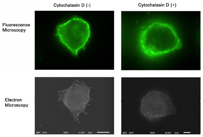
Photo by Andor Technology plc.
CD44 is recognised as an important cell surface receptor involved in the adhesion of circulating leukocytes to endothelium, a process that is critical for their trafficking in the vasculature. Now, a Japanese group of researchers using an ultra-sensitive Andor Neo sCMOS camera in their correlative microscopy set-up has demonstrated that microvilli projections around the cell surface mediate this adhesion.
Although fluorescence imaging is a powerful approach to studying the cellular distribution of membrane receptors, it is limited in its ability to display fine structural information. Therefore, the team mapped the distribution of CD44 at the lymphocyte cell surface in liquid by correlative immune-fluorescence optical and immuno-electron microscopy using a combined fluorescence and atmospheric scanning electron microscope. Once CD44 cell surface labelling was confirmed with the images from the Neo sCMOS detector, atmospheric SEM revealed the microvilli on the cell surface with the localisation of label on the microvilli.
“Dual labelling was employed using an anti-CD44 monoclonal antibody and a secondary antibody conjugated with Alexa Fluor 488 and 1.4 nm positively charged Nanogold particles,” says Toshiyuki Murai, lead author of the paper. “We also demonstrated that treatment of cells with cytochalasin D resulted in the loss of the microvilli projections and the simultaneous nullification of CD44-mediated adhesion to its ligand hyaluronan. These results suggest the functional relevance of microvilli in CD44-mediated rolling adhesion under shear flow.”
The correlative light and SEM imaging was accomplished by culturing the mouse lymphocytes in a petri dish to which a 100 nm silicon nitride window was fixed. This window is transparent to the electron beam, which is projected from beneath the dish by the inverted SEM, while the upright fluorescence microscope excites the sample from above through the objective lens.
“Atmospheric or Environmental SEM enables life scientists to examine biological specimens in exquisite detail, far beyond the optical diffraction limits associated with optical microscopy,” says Orla Hanrahan, product specialist at Andor. “It can be used to observe mammalian cells, bacteria, and protein crystals, for instance, and its impact in correlative microscopy studies of cellular structure and function cannot be underestimated. For these studies, Andor’s Neo 5.5 megapixel sCMOS is the perfect, ultra-sensitive, high-resolution partner for fluorescence imaging.”
Andor’s Neo 5.5 megapixel sCMOS camera is a unique -40°C vacuum cooled platform to drive lowest possible dark noise. Neo 5.5 has low noise and excellent resolution with a 5.5 megapixel sensor with 6.5 µm pixels and a 22 mm diameter. Ideal for cell microscopy, astronomy, digital pathology, and high content screening, Neo 5.5 delivers an unmatched 30 fps sustained or up to 100 fps burst mode to its internal 4 GB memory. The Rolling and Global shutter flexibility further enhances application flexibility, Global shutter in particular offering an ideal means to simply and efficiently synchronize the Neo with other ‘moving’ devices such as stages or light switching sources and eliminating the possibility of spatial distortion when imaging fast moving objects.
For more information, please visit http://www.andor.com.
News Categories
- » NEWS HOME
- » Automation & Robotics
- » Industry 4.0
- » Material Handling
- » Sensors
- » Quality & Testing
- » Machine Vision
- » Laser & Optics
- » Metalworking
- » Motion Control & Drives
- » Hydraulics & Pneumatics
- » Process Industry
- » Renewable Energy
- » Agriculture
- » Home & Office Furniture
- » Environmental Tech

Tags
|
|
|
|
|
|
|
|
|
|
|
|
|
|
|
|
|
|